


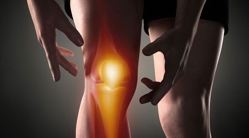
Knee hurts- This is a sign of a pathological process affecting the cartilage, bone or soft tissue structure of the femur - tibia and femur. Joint pain can be based on trauma, inflammatory and degenerative diseases of the articular apparatus and periarticular structures. Patients may complain of sharp, aching, burning, throbbing, and other types of pain that occur at rest or when moving, supporting, flexing, and extending the leg at the knee. Diagnosis of causal pathology includes instrumental imaging methods (Rg, ultrasonography, CT or MRI, arthroscopy), joint capsule puncture, biochemical and immunological analysis. Until the diagnosis is clarified, rest, joint immobilization, NSAIDs, and analgesics are recommended.
Causes of knee pain
Injury
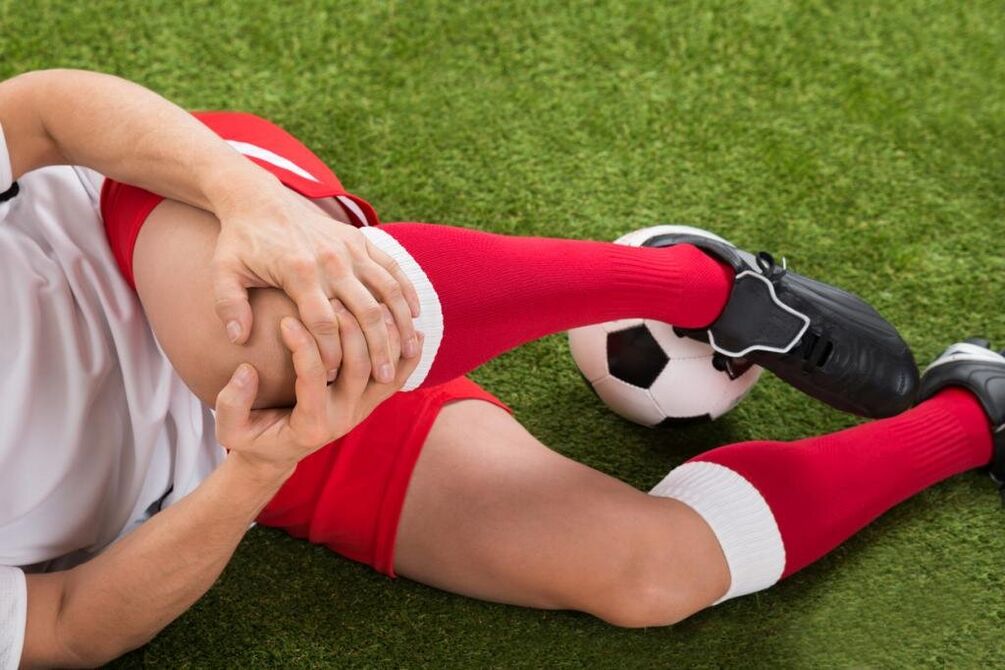
Most often they are the result of family trauma, often seen in athletes: runners, diving athletes, sports participants. Developed by a fall, direct impact, or twisted leg. Manifested by sharp pain at the time of injury. In the future, the pain syndrome becomes less pronounced, accompanied by increasing edema. May be scratched and bruised. As frequency increased, the following injuries were identified:
- Knee Injury. . . Occurs when falling to the knee or hitting it directly. At first, the pain is sharp, hot, sometimes burning, but tolerable, then - a dull, aching pain that worsens with movement. There may be bruising. Support legs are kept. Sometimes knee injuries are complicated by joint sequelae, in such cases the joint gradually increases in volume, becomes spherical, a feeling of pressure or ruptures is added to the pain syndrome.
- Ligament rupture.It is found after twisting, twisting, bending, or overexerting in a non-physiological position. Pain is stronger than a bruise; Simultaneously with the onset of pain, a person may feel something tearing (similar to how normal tissue is torn). Accompanying it is a significant limitation in movement, support, limb contortion, rapid increase in the rate of metastasis.
- Fractures in joints. . . They are detected during collisions, falls and twists. In the event of an injury, a person feels a sharp, often unbearable pain, sometimes hearing a crunching sound. Patients with intra-articular fractures themselves describe their feelings as follows: "The eye hurts so much that it goes dark, the world doesn't exist, you don't understand anything". After that, the pain becomes less severe, but still intense. Support is often impossible, movement is almost completely restricted. Edema and metastasis progress rapidly.
- Dislocation.As a result of a blow or fall to the knee. At the time of dislocation of the kneecap, the patient presents with sharp pain, accompanied by a feeling of bowing and the knee joint is displaced. Can't move, can save function reference. On the anterior surface of the knee, a pronounced deformity is visible, which is subsequently smoothed by increasing edema. Sometimes hereditary hereditary infections (hemarthrosis) are involved.
- Pathological fracture. They develop with minor trauma, as a result of reduced bone strength in osteoporosis, osteomyelitis, tuberculosis, osteosarcoma. Aching, dull ache, reminiscent of pain syndrome with bruising. Signs of a pathological fracture include limited or no support in the leg, a feeling of instability in the knee, sometimes deformity, and a crunching bone with movement.
- Damage to the menisci.Meniscal tears are formed during intense twisting, impacting, flexion, or extension of the knee with a fixed leg. At first, a person feels a special click and sharp pain deep in the joint. After that, the pain subsides somewhat, but becomes diffuse, sometimes - burning, flare, more intense when trying to prop up and move. The mass of the knee increases due to edema and metastasis. Support becomes impossible, movements are strongly restricted.
Inflammatory disease
They can be infectious and non-infectious (post-traumatic, toxic, metabolic, post-vaccination). Abundant blood supply to the synovial membrane and periarticular tissues promotes rapid development of inflammation in response to direct and indirect effects, and a large number of nerve endings induce a pronounced pain response. . The inflammatory process is often accompanied by bursitis (accumulation of sterile fluid in the joint), infection, possible accumulation of pus.
- Arthritis.Inflammation of the gonads occurring after trauma, sometimes complicating infectious diseases, is found in rheumatic diseases. Can be acute or chronic. Knee pain is often dull, aching, pressing or pulling. At first, the pain was not intense and intermittent, increasing gradually in the evening or after exercise. Then the pain begins to join, the intensity and duration of the pain syndrome increases. Joints swell, skin reddens, temperature rises. With bursitis, the contours of the knee are smooth, there is a feeling of tightness. When the pain is reduced, the severity of the pain increases sharply, they become convulsive, lose sleep.
- Bursitis.It is not an independent disease, a complication of many acute and chronic pathologies of the joints. It is formed within hours or days. Initially, the pain is insignificant or absent, and satiety predominates. The knee is spherical, the fluid is abundant, the skin is glossy. Movement is somewhat limited. When infected, the pain becomes pronounced, vibrating, convulsing, more intense with even the slightest movement and touch.
- Bursitis.Inflammation of the patella and patella commonly occurs when the knee is overloaded and repetitively traumatized (for example, when the knee is supported continuously). With bursitis, local pain, dull, not intense, appears at a certain position of the limb, which, after bearing a characteristic force, decreases with changing the position of the leg, massaging the affected area. If the posterior sac is affected, there may be pain when walking up or down stairs. Small local edema is sometimes identified. With the cessation of the pain, the pain becomes sharp, convulsions, grilling, combined with congestion, edema of the affected area, symptoms of general intoxication.
- Tendonitis.Usually found in overweight men and athletes, it affects the ligaments of the patella. Initially, pain syndrome occurs only with very vigorous exertion, then with standard sports loads, then with daily physical activity or at rest. Tendonitis pain localized anteriorly just below the knee, dull, tugging, progressive, sometimes paroxysmal, in some cases accompanied by mild redness and swelling that worsens with pressure. Movement is usually total, less often slightly restricted. There may be a tear or rupture of a ligament due to decreased strength of the ligament.
- Fat inflammation.Hoff's disease affects the fatty tissue layers below the kneecap. It is observed with constant overload of the knee or becomes the result of an old injury. More often it affects athletes, older women. A person complains of dull pains combined with some limitation of prolongation. With the exacerbation of the pathology, the pain begins to disturb at night, there is a feeling of unsteadiness in the knee, and the legs are bent. When the side of the kneecap is pressed, a cracking or creaking sound may be heard.
Autoimmune process
The cause of diseases of this group is the production of antibodies to the normal cells of the body with the development of aseptic immunological inflammation of the synovial membrane and cartilage, inflammation of the blood vessels. The diseases are in most cases chronic, if left untreated, they are easily progressive and are often the cause of disability.
- Rheumatoid arthritis.Success and failure are often bilateral. With minimal activity of the autoimmune process, the pain is weak or moderate, intermittent, pulling, pressing, accompanied by morning stiffness. With moderate activity, the patient complains of persistent, pressing or recurring pain of moderate intensity, not only during exercise but also at rest. There is stiffness for many hours, moderate recurrent bursitis. With the high activity of rheumatoid arthritis, the pain is strong, diffuse, tired, of a rippling nature, which increases in the hours before morning. The tension becomes constant, large amounts of fluid accumulate in the knee, and spasticity develops over time.
- Systemic lupus erythematosus.Occlusions are usually symmetrical, although one joint may be affected. They can occur at any stage of the disease; with a recurrent episode of SLE, they resemble rheumatoid arthritis. With low process activity, the pain is short-term, not intense, localized, aching, pulling. In severe cases, the pain syndrome progresses, pain ripples, causes nocturnal sleep disturbances, lasts, diffuses, increases with movement, associated with bursitis, edema, and congestion.
- Rheumatism.Joint pain is one of the first manifestations of rheumatic fever, appearing 5-15 days after an acute infection, affecting multiple joints at once (usually in pairs). The pain is short-lived, but intense, moves from one joint to another, and varies in nature from pulling or pressing to burning or vibrating. Knees are swollen, burning, and the outer skin is red. Movement is severely limited. After a few days, the pain level subsides, movement is restored. In some patients, residual effects in the form of moderate or mild dull pain persist for a long time.
- Reactive arthritis.Usually occurs more than 2-4 weeks after intestinal and urogenital infections, usually affecting one or two joints of the lower extremities, associated with urethritis, conjunctivitis. The development of reactive arthritis precedes increased urination, pain and burning sensation in the urethra, tearing and cramping in the eyes. Severe or moderate pain in the knee, continuous, rippled, aching, tugging, convulsing, associated with limitation of movement, worsening of general condition, fever, severe swelling and redness of the affected area. Pain and signs of inflammation last from 3 months to 1 year, then gradually disappear.
Degenerative-dystrophic processes
They develop due to metabolic disturbances in the structure of the joints and the soft tissues around the joints. They have a chronic, progressive course over many years. Often accompanied by the formation of calcifications, cysts and osteomas, and deformation of the knee surface. With significant destruction of articular surfaces, they lead to marked impairment of motor and support function, becoming a cause of disability and requiring intracellular installation.
- Osteoarthritis.It develops for no apparent reason or against the background of various injuries and illnesses, mainly in the elderly and middle-aged. Initially, short-term weakness, often pulling or aching, occurs with prolonged exertion and disappears with rest, often accompanied by a crunching sound. Gradually, the pain syndrome increased, the knee began to ache "because of the weather" and at night, limited movement. Distinctive features of gonarthrosis are the onset of pain (pain until you "dissolve"), periodic attacks of pain such as cutting, burning, or shooting from the blockade. During the exacerbation phase, bursitis often occurs, in which the pain becomes constant, pressing, exacerbating.
- Meniscopathy. . . Usually found in athletes, whose work involves a significant load on the knee joint. Presented by local deep pain on one side of the knee at the joint space level, more often in the outer half of the knee. The pain increases with movement and subsides with rest and may be dull, pressing, or pulling. With progression, there is acute pain when trying to move. On the anterior lateral surface of the joint when pain occurs, a small pain can sometimes be felt.
- Tendon disease. . . The tendons near the knee are affected. In the early stages, they are manifested by short-term localized superficial pain at the peak of physical activity. Then pain occurs with moderate loads, and then with light loads, limiting usual daily activities. Traction pain or tenderness, directly related to active movements, is not detected with passive extension and flexion of the knee, sometimes accompanied by a crunching or crunching sound. In the affected area, the most painful site can be probed. Local inflammatory markers (edema, congestion, hyperthermia) were insignificant or absent.
- Bone disease.Children and young people are more often affected, the duration of the disease is several years. Usually, they begin gradually with mild lameness or intermittent dull pain, not intense, worsen with exertion, disappearing with rest. With the progression of osteosarcoma, the pain becomes strong, constant, pressing, burning or toasting, accompanied by severe lameness, limited range of motion, and difficulty resting in the extremities. After that, the pain subsides and the supporting function is restored.
- Chondromatosis.Usually diagnosed in older men, less often in infants. Arthritis chorioamnionitis is manifested by moderate dull pain, which often worsens at night and in the morning. Movement is limited, accompanied by a crunching sound. Obstruction sometimes occurs, characterized by sudden sharp pain, inability or severe limitation of movement. With the development of bursitis, the pain has a flare-up nature, combined with an increase in the volume of the knee, swelling of the soft tissues and an increase in local temperature.
Tumor and tumor-like formation
Pain syndrome can be caused by cysts, benign or malignant tumors that directly affect joint tissues or peristalsis. In addition, knee pain can be a warning sign of hypertrophic joint disease, pancreatitis polyarthritis - a paraplastic syndrome characteristic of lung cancer, breast cancer and other cancer processes.
- Baker's cyst.Represents skull protrusion in popliteal fossils. In the early stages, it manifests as local discomfort or mild pain along the back of the knee. Against the background of an increase in a Baker's cyst due to compression of adjacent nerves, a single area of burning or burning pain, numbness or tingling may occur. Symptoms are worse when trying to bend the knee as much as possible. Occasionally, an elastic, slightly painful lump-like mass may be felt.
- Benign tumors.These include chondromas, osteosarcomas, undifferentiated fibroids, and other tumours. They are characterized by a prolonged period of no or few symptoms, which may manifest themselves as localized pain that is not intense and vague. With large tumors, solid formations can be felt, sometimes developing synovitis.
- Malignant tumor.The most common malignancies affecting the joint area are synovial sarcoma, osteosarcoma, and chondrosarcoma sarcoma. They manifest as local dull ache, sometimes following a certain circadian rhythm (worse at night). The intensity of the pain increases, they become sharp, cutting, burning or convulsing, spreading along the knee and adjacent tissues, accompanied by deformity, edema, synovitis, dilation of the veinshemisphere, violation of the general condition, the formation of shrinkage. Palpation identifies a painful tumor-like formation. Once the process begins, the pain is intense, intolerable, exhausting, causing you to lose sleep and not be eliminated by non-narcotic pain relievers.
Invasive operations and manipulations
Pain syndrome is triggered by damage to knee tissue during invasive procedures. The severity of the pain directly depends on the injury of the manipulation on the knee joint. With the penetration of pathogenic microorganisms into the joint area, pain is caused by inflammatory changes.
- Operation.The most common procedure is puncture. Post-puncture pain is short-lived, not intense, rapidly subsides, localized to the projection of the puncture, usually performed on the lateral aspect of the knee. After the biopsy, the pain may be twitchy at first, then dull and disappear after a few days.
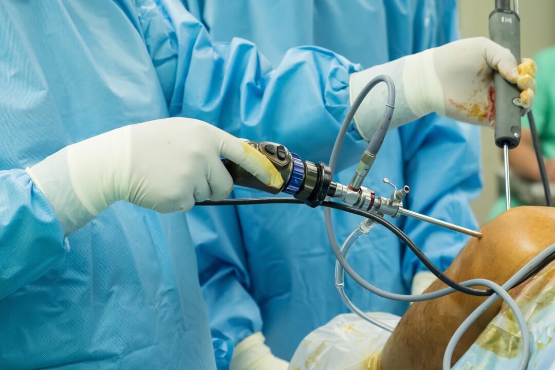
- Activities.After arthroscopy, the pain is moderate, rather acute at first, then dull, which subsides after a few days or 1-2 weeks. After joint removal surgery, the pain is more intense, which can last up to several weeks due to significant tissue damage. Usually, in the first 2-3 days after the intervention, the patient is prescribed pain medication, then the pain gradually weakens and disappears.
Psychological conditions
Sometimes joint pain in the knee occurs without an organic basis (injury, inflammation, destruction, . . . ) under the influence of psychological factors. Such pain is believed to play a protective role, as it helps reduce emotional stress by converting the experience into a physical sensation. The distinguishing feature of such pains is their indeterminate nature, inconsistency, absence of visible changes, clear association with physical activity and triggers. other objective. Deformed arthropathy is observed in people who are sensitive to changes in barometric pressure.
In addition, radiation therapy for knee pain can occur with coxarthrosis, lumbar osteochondrosis, Perthes disease, fibromyalgia, sciatica. However, with these diseases, pain syndromes of other loci often appear first. Additional risk factors that increase the likelihood of knee injury and disease include excessive weight, professional sports, anemia, metabolic disorders, and old age. Hypothermia, stress, exercise, and dietary disturbances can all be triggers that exacerbate chronic pain.
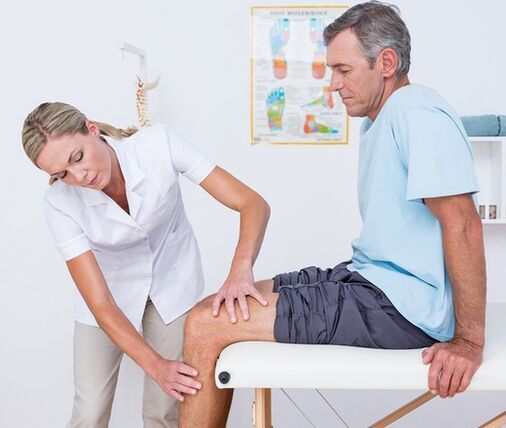
survey
The algorithm searches for a diagnosis based on taking into account the nature of the pain syndrome, its duration, and identifying concomitant symptoms and events prior to the onset of knee pain. At the first visit with the doctor (trauma-orthopedist, surgeon, rheumatologist), an imaging examination and palpation of the knee, assessment of the volume of voluntary movementsand passivity is performed. Taking into account the data obtained, in the future the patient can be assigned:
- Laboratory blood test. . . A complete blood count helps to identify hematological changes characteristic of an acute inflammatory and infectious process (leukocytosis, increased ESR), eosinophilia, typical of an allergic reaction. Biochemical and serological studies are the most informative for autoimmune diseases, which are characterized by the formation of specific acute-phase proteins and immunoglobulins (CRP, rheumatoid factor, ASL-O, CEC, antibodies to DNA, etc. ).
- X-ray.The basic diagnostic method is X-ray of the knee joint according to 2 projections. The presence of pathology is indicated by changes in the contour of the head and joint cavity, narrowing of the joint space, changes in the thickness of the end plates, the presence of marginal defects at the joint ends, resolutionbone and bone destruction. . In some diseases (injury to meniscus, Baker's cyst), contrast arthrography exhibits the greatest sensitivity.
- Arthrosonography. . . Knee ultrasound is a quick, inexpensive, affordable, and highly informative diagnostic method. Allows you to evaluate the presence of effusion and free bodies in the joint cavity, to identify lesions and pathological changes in the periarticular soft tissues (signs of calcification, hemorrhage, etc. ). They help to distinguish with high accuracy the etiology of joint pain.
- CT and MRI. . . They are the methods of choice for joint disease of any origin. They are used for a more detailed assessment of the nature and extent of pathological changes, identifying typical signs of traumatic, inflammatory, and neoplastic lesions of bone and soft tissue structures. Joint CT and MRI are often used with the limited informational content of other instrumental studies.
- Joint perforation. . . It is performed when there is evidence of accumulation of exudate or fluid in the joint capsule. As part of the differential diagnosis of inflammatory, degenerative and neoplastic diseases, a cytological, bacteriological or immunological study of the synovial fluid is performed. To confirm the diagnosis of autoimmune knee joint injury, tuberculosis arthritis, synovial tumor, conducting a biopsy of the synovial membrane is extremely important.
- Arthroscopy. . . The aim of invasive endoscopic diagnosis may be to obtain a biopsy sample, clarifying the diagnostic information needed during visual examination of joint elements. In some cases, diagnostic arthroscopy evolves into a treatment (arthroscopic resection of intra-articular tissues, meniscalectomy, autologous angioplasty, etc. ).
Symptomatic treatment
Treatment of the causes of knee pain is carried out differently, taking into account the identified disease. At the same time, symptomatic care is an essential part of a comprehensive treatment that aims to reduce discomfort and improve quality of life. Immediately after an injury, apply a cold compress to the knee area - this will help reduce pain sensitivity. Ethyl chloride has a cooling and local anesthetic effect. In any case, resting the knee will help reduce soreness. It is necessary to limit movement, put the leg in the position with the least pain. When walking, a fixed bandage is applied to the knee, which can be immobilized with the help of a plaster cast.
In the acute stage of injury or illness, absolutely do not massage your knees, apply warm compresses, or wear high heels. The main classes of drugs used to treat pain and inflammation are pain relievers and NSAIDs in the form of ointments, tablets, and injections. The remedies listed can only provide temporary relief, but cannot eliminate the root cause of joint pain. Therefore, all cases of knee pain require a qualified diagnosis and treatment, and some conditions (fractures, dislocations, metastases) require urgent medical attention. You cannot postpone a visit to the doctor if the pain is associated with a change in the shape of the knee (swelling, smoothing of contours, asymmetry), inability to perform stretching, patellar movement, impaired ability of the limb to support.